診察時間
午前9:00-12:00
午後15:00-19:00
手術時間12:00-15:00
水曜・土日午後休診

尿管結石🐈💎
-
-
尿管結石とその影響
尿管に結石ができると腎臓の構造が変化し、本来排出されるべき尿毒素が体内に蓄積し、急性腎不全(腎臓が突然正常に機能しなくなる状態のこと)へと進行し致死的な状態になることがあります。腎数値の上昇時には、エコー検査で尿管や腎臓に結石がないかを確認し、急性腎不全と慢性腎不全の急性転化を区別することが重要です。
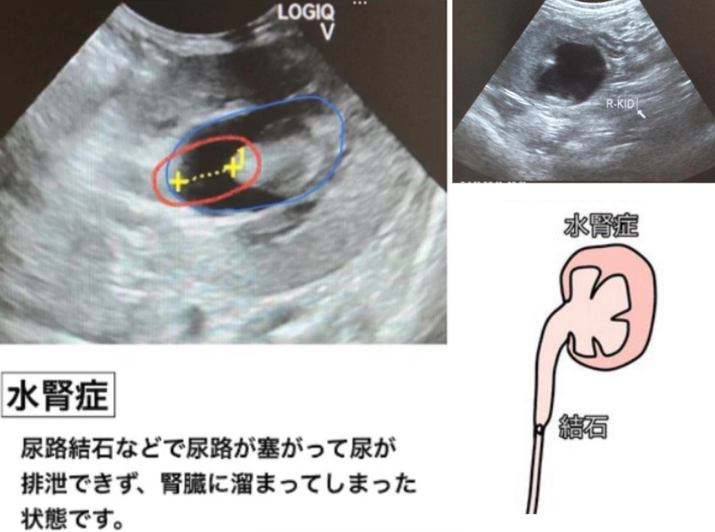
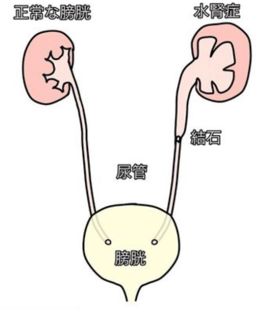
尿管結石は腎臓から膀胱へ尿を送る尿管に詰まり、腎臓に尿が溜まり水腎症を引き起こし、最終的に腎臓の壊死につながります。特にねこちゃんに多い疾患です。
-
-
診断と治療計画
反対側の腎臓が代償している間は、排尿は通常通りであり、腎臓を触ると痛みを感じることがありますが、臨床症状だけでは早期診断は難しいです。レントゲンやエコー検査で腎臓の構造、腎盂や尿管の拡張、結石の有無を確認し、手術計画を立てます。
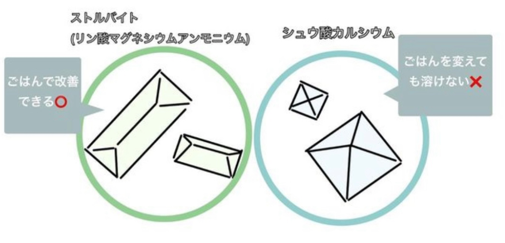
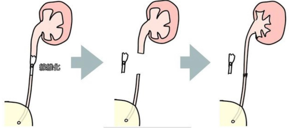
シュウ酸カルシウムのように療法食で治療不可能な結石がある場合、特に細い尿道を閉塞している場合は結石を摘出する必要があります。尿管の構造が線維化し、尿の通過機能を損なうと、石を摘出しても尿が排出されないことがあります。その場合、異常な尿管を切除して膀胱に繋ぐ新尿管膀胱吻合術が必要です。
-
-
外科手術の詳細
外科手術では、尿管の閉塞している箇所を切開し、結石を取り除きます。閉塞していた期間が長いと尿管が線維化しており、その部分は切除し正常な尿管を繋ぎ合わせる必要があります。
手術に際しては、おへそから骨盤前方まで大きく開腹し、消化管や腹膜内脂肪を避け、隣接する大血管に注意しながら尿管と腎臓を露出させます。尿管を触診して結石の位置を特定し、結石を摘出後は尿管を丁寧に縫合し、尿漏れがないことを確認して閉腹します。

-
-
手術後のケアと注意点
術後は点滴療法での入院治療を行い、縫合部位の離開や心不全がなく、元気と食欲が安定していれば退院となります。腎臓内にも結石がある場合には、定期的にエコーや血液検査で尿管の閉塞がないかを確認します。
非常に繊細な手術であるため、縫合箇所からの尿漏れや、急性腎不全が生じている場合には腹膜透析を行い、毒素を除去します。また、尿管が狭窄しないように、粘膜の切開・縫合箇所の腫れにも注意が必要です。
-
-
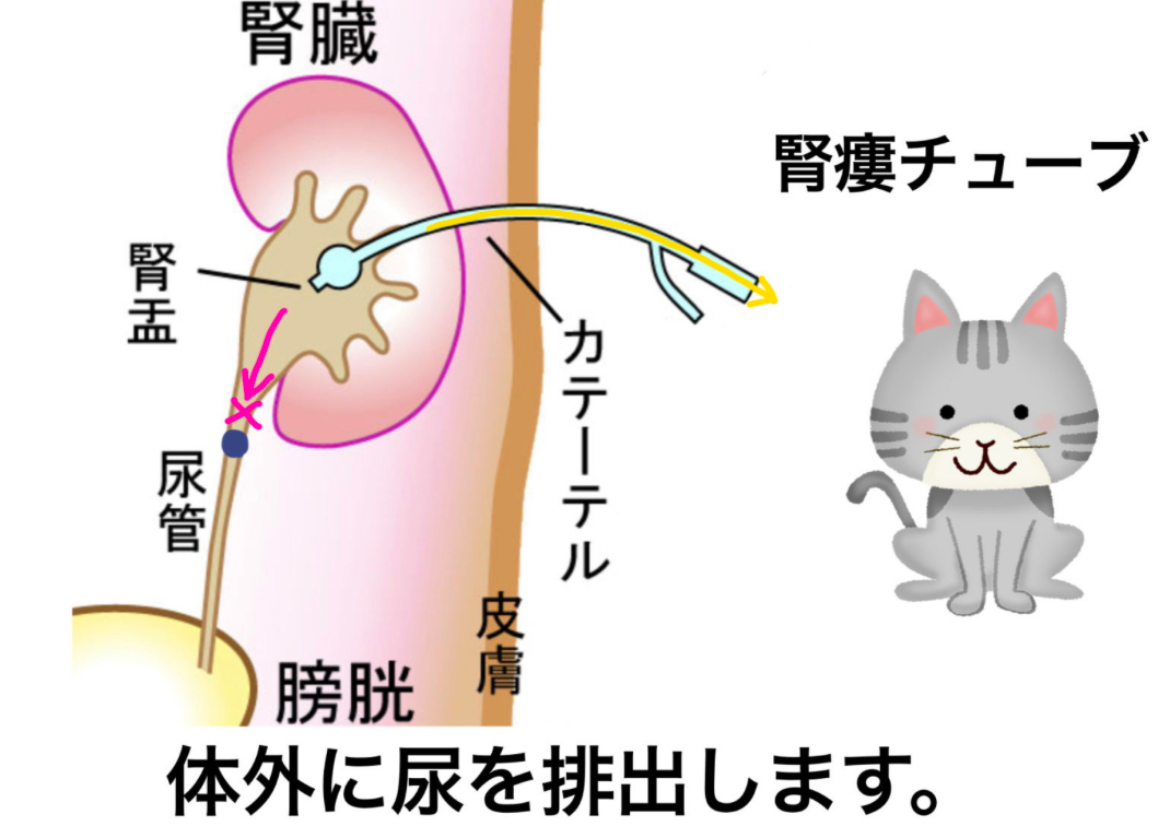
水腎症に対する腎瘻チューブの使用
水腎症やその他の尿路閉塞症状が見られる場合、術前の状態を安定化させるために腎瘻チューブが使用されます。これは、尿管結石によって引き起こされる尿の停滞と腎臓への圧迫を緩和し、急性腎不全を防ぐための一時的な措置です。腎瘻チューブの挿入は、麻酔をかける前にペットの状態を最適化し、手術への耐性を高めることを目的としています。
この処置により、手術時のリスクが軽減され、手術後の回復にも有利に働きます。腎瘻チューブは、腎臓から直接尿を外部に排出するため、尿路の圧力を減少させ、腎機能の一部を保護します。
-
-
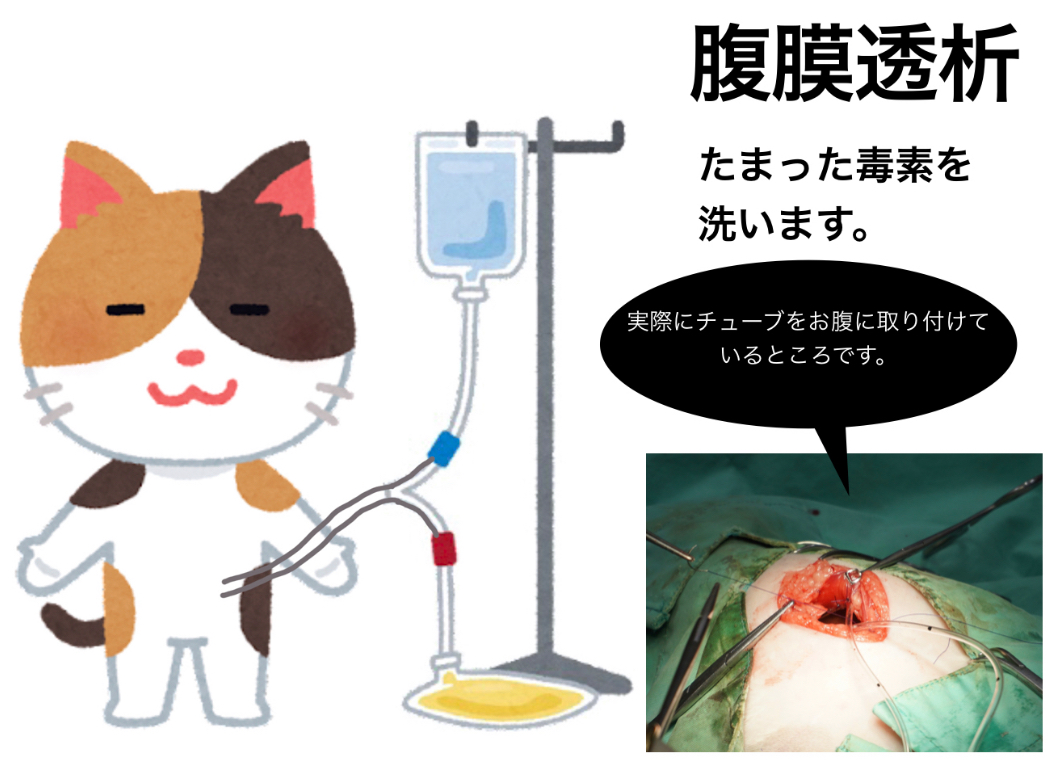
尿管結石摘出後の腹膜透析
尿管結石を摘出する手術後、特に腎機能が低下している場合には、腹膜透析を行うことがあります。この追加の治療は、麻酔からの回復中に、体内に蓄積された毒素を効果的に除去するために行われます。
腹膜透析は、腹膜をフィルターとして使用し、血液中の毒素や余分な水分を体外に排出する方法です。手術によって短期間に腎機能が低下した場合や、急性腎不全のリスクが高いペットにとって、生命を維持する重要な支援措置となります。