

診察時間
午前9:00-12:00
午後15:00-18:00
手術時間12:00-15:00
水曜・日曜午後休診
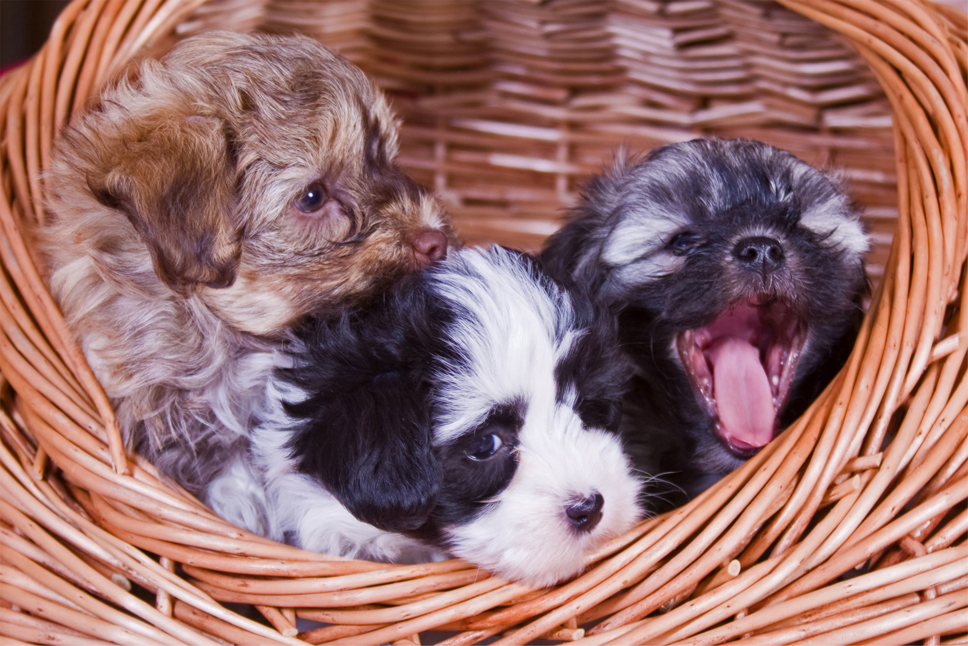
犬の上気道疾患と手術・麻酔管理のやさしい解説|短頭種気道症候群・喉頭麻痺
上部気道の病気と手術・麻酔の前後の管理について、やさしく解説します
こんな様子、思い当たりませんか?
・いびきが大きく、寝ているときに「ガーガー」と音がする
・運動するとすぐにゼーゼーして座り込んでしまう
・興奮したり暑くなると、苦しそうにハアハアして舌が紫っぽくなる
・吐き気や嘔吐が多い、食後にむせることがある
こうした症状の裏側には、上部気道(鼻〜咽頭〜喉頭)の病気が隠れていることがあります。
代表的なのが短頭種気道症候群や喉頭麻痺などです。

Ⅰ.上部気道疾患とは?
「上部気道」とは、鼻腔・咽頭・喉頭・気管の入り口までの空気の通り道を指します。
このどこかが狭くなったり、柔らかい組織が気道内にたれ込んだりすると、
空気の流れが悪くなり、次のような症状が現れます。
- 大きないびき、睡眠時の「ガーガー」という音
- 運動時・興奮時のゼーゼーした呼吸(努力呼吸)
- 口を大きく開けてハアハアする、呼吸が速く浅い
- 舌や歯ぐきが紫っぽくなる(チアノーゼ)
- 暑い日に突然倒れそうになる、失神してしまう
- 吐き気・嘔吐、食後のむせやすさ など
代表的な上部気道疾患には、次のようなものがあります。
- 短頭種気道症候群(brachycephalic airway syndrome:BAS)
- 喉頭麻痺(声帯を動かす神経の麻痺)
- 喉頭小嚢外反・喉頭虚脱
- 気管虚脱や咽頭・喉頭・気管の腫瘍 など
上部気道疾患の怖いところは、興奮・暑さ・ストレスをきっかけに一気に状態が悪化し、命に関わる呼吸困難に進むことがある点です。
そのため、病気そのものの治療だけでなく、手術の前後(周術期)の呼吸管理がとても大切になります。
Ⅱ.呼吸状態の評価と「換気」と「酸素化」
呼吸の状態を見るときには、「換気」と「酸素化」を分けて考えます。
- 換気:息をしっかり吐いて、体の中の二酸化炭素(CO₂)を外に出せているか
- 酸素化:吸い込んだ酸素(O₂)がきちんと血液に取り込まれているか
1.動脈血ガス分析で見る項目
血液ガス検査では、主に次の値を確認します。
- pH:血液の酸性・アルカリ性のバランス
- PaCO₂:動脈血中の二酸化炭素分圧(換気の指標)
- PaO₂:動脈血中の酸素分圧(酸素化の指標)
- HCO₃⁻・BE:代謝性の補正の状態 など
犬猫で FiO₂ 0.21(通常の空気)を吸っているときの、おおよその目安は以下の通りです。
- PaCO₂
・約 35〜45 mmHg:ほぼ正常域
・それ以上:換気低下(呼吸が足りず CO₂ がたまっている)
・大きく低下:過換気(逆に呼吸しすぎている) - PaO₂
・80 mmHg 以上:ほぼ正常
・60〜80 mmHg:軽度の低酸素血症
・60 mmHg 未満:明らかな低酸素血症で要注意
2.非侵襲的モニタリング(SpO₂・ETCO₂)
- SpO₂(パルスオキシメータ)
指や舌につけるクリップ型の機械で、血液中の酸素飽和度を測ります。
・95〜100%:十分な酸素化
・90%前後:注意の必要なゾーン
・90%未満:低酸素血症を疑う値 - ETCO₂(カプノメータ)
「吐き終わりの息」の CO₂ 濃度をリアルタイムで測定します。血液ガスの PaCO₂ に近い値となるため、
換気状態の変化や麻酔中のトラブルの早期発見にとても役立ちます。
Ⅲ.酸素療法の方法と注意点
上部気道疾患の子では、症状が強いときに酸素を補ってあげることがとても大切です。
状態や性格に合わせて、いくつかの方法を使い分けます。
- フロー・バイ(flow-by)
顔の前から酸素を流す方法です。拘束が少なく短時間の応急処置に向きますが、取り込める酸素濃度は 30〜40%程度までです。 - フェイスマスク
マスクで口や鼻をおおい、より高い酸素濃度(40〜70%)を保てます。
ただし、苦手な子ではかえって興奮やストレスで呼吸が悪くなることがあります。 - 経鼻カテーテル
鼻の中から細いチューブを挿入して酸素を流す方法です。比較的自由に動けるため、長時間の酸素投与に向きます。 - 気管内カテーテル(挿管中)
麻酔中など、気管チューブが入っている状態では、確実に気道を確保しながら高濃度酸素を送ることができます。 - 酸素ケージ
密閉されたケージ内の酸素濃度を上げる方法です。
興奮しやすい子でもあまり触らずに管理できる点がメリットですが、扉の開閉で酸素濃度が変動しやすく、
温度・湿度の管理も必要になります。
高濃度酸素の“やりすぎ”にも注意
- 長時間の高濃度酸素(特に FiO₂ > 60%)は、肺の細胞を傷つける「酸素毒性」の原因になることがあります。
- 窒素が少ないガスを吸い続けると、肺胞がしぼんでしまう「吸収性無気肺」が起こり、かえってガス交換が悪くなることがあります。
- 加湿されていない酸素を長時間流すと、気道粘膜が乾燥し、分泌物が粘ついて詰まりやすくなります。
そのため、「必要なときに、必要な量を、できるだけ短期間に」
という考え方で酸素療法を行うことが大切です。長期にわたる場合は、FiO₂ を 50%以下に抑えたり、加湿を併用したりといった工夫を行います。
Ⅳ.手術前後(周術期)の全体的な流れ
上部気道疾患の子では、手術そのものと同じくらい、麻酔の前後の管理が重要です。
ここでは一般的な流れをご紹介します。
1.手術前の安定化
- 興奮・ストレス・暑さをできるだけ避け、静かで涼しい環境で休ませる。
- 必要に応じて酸素投与を行い、SpO₂ や呼吸の様子を確認する。
- 重度例では動脈血ガスを測定し、換気・酸素化の程度を客観的に把握する。
- 不安の強い子では、慎重に選んだ鎮静薬で落ち着かせ、かえって呼吸が楽になるよう配慮する。
2.麻酔導入時の注意点
- 麻酔薬を投与してから気管挿管が完了するまでの時間をできるだけ短くする。
- 複数サイズのチューブ・スタイレット・ビデオ喉頭鏡などを準備し、「挿管しにくい場合の次の一手」をあらかじめ決めておく。
- 最悪の場合に備えて、緊急気管切開の器具も手元に用意しておく。
3.術中管理
手術中は SpO₂・ETCO₂・心電図・血圧・体温などを連続モニタし、数値の変化から早めにトラブルをキャッチします。
ETCO₂ の上昇や SpO₂ の低下が見られたときには、換気量の調整・チューブ位置の確認・喀痰吸引などを行います。
4.覚醒・抜管時が最大の山場
上部気道の腫れが残っていたり、痛みや不安で暴れてしまったりすると、
抜管後に急激な呼吸困難を起こす危険があります。
そのため、次のような状態を確認してから抜管します。
- 自力でしっかり頭を持ち上げられる
- 舌を引っ込めたり、飲み込む動きができる
- 呼吸が落ち着いている(努力呼吸がない)
短頭種や重度症例では、通常より長く挿管を続け、完全に覚醒してから抜管することも多いです。
抜管後もしばらく酸素を続け、静かな環境で慎重に観察します。
Ⅴ.短頭種気道症候群(BAS)の病態
フレンチブルドッグ、パグ、ボストンテリア、キャバリアなどの短頭種は、
生まれつき顔の骨格が短く、のどや鼻の構造に負担がかかりやすい犬種です。
短頭種気道症候群では、次のような一次性病変と二次性病変が組み合わさって進行していきます。
- 一次性病変
・外鼻孔狭窄(鼻の穴が細い)
・軟口蓋過長(のどの奥の柔らかい天井が長すぎる)
・喉頭小嚢外反
・低形成気管(気管の直径が細い)など - 二次性病変
長期間の強い努力呼吸により、喉頭や気管が潰れてくる喉頭虚脱・気管虚脱、肺への負担増加などが起きます。
「いびきがひどい」「散歩が昔より苦しそう」といったサインは、
こうした変化が少しずつ進んできている合図かもしれません。
早めに介入するほど、二次性病変が出る前に負の連鎖を断ち切りやすくなります。
短頭種気道症候群の重症度分類
文献では、いびきの頻度・運動時の呼吸困難・睡眠時の無呼吸・チアノーゼや失神の有無などをスコア化し、
0〜4 点程度の重症度グレードで評価しています。
一般的には、
- グレード 0〜1:軽度(生活上の工夫と経過観察中心)
- グレード 2〜3:中等度(外科手術を積極的に検討)
- グレード 4:重度(できるだけ早期の外科介入を考慮)
上部消化管疾患との関連
短頭種気道症候群の子では、胃食道逆流や胃炎、幽門狭窄、胃内容停滞などの消化器疾患を併発しやすいことがわかっています。
強い陰圧が長く続くことで、胃や食道の内容物が逆流しやすくなるためです。
手術前後には、制酸薬・胃運動改善薬などの内科治療、絶食時間の調整、術後の食事の与え方の工夫が重要になります。
Ⅵ.外鼻孔狭窄の構造と手術
外鼻孔(鼻の穴)の周囲は、鼻翼や外鼻孔翼軟骨などの柔らかい組織で構成されています。
短頭種では、これらが内側に折れ込むように発達してしまい、穴の入口が物理的にふさがっている状態になっていることがあります。
吸気時には外側の組織がさらに内側へ吸い込まれ、空気の通り道がますます細くなります。
このため、外鼻孔を外側に開いてあげる手術が重要になります。
代表的な手術法
- 鼻翼楔状切除術(垂直楔形切除)
鼻翼の一部をメスで楔形に切除し、残った辺縁を外側へ引き出すように縫合する方法です。
切除量を調整しやすく、幅広く行われています。 - パンチ切除術
鼻翼基部にパンチ(2〜6 mm)を打ち、円形に組織を切り取ります。
その後、穴の縁を外側へ開くように縫合します。左右差が少なく、仕上がりが整いやすい方法です。 - 鼻翼固定術
鼻翼の外側を部分的に切開し、外側へ引き出した位置で皮膚と軟骨を縫合して固定する方法です。
ほかの手術と組み合わせて行うこともあります。
いずれの方法でも、「必要なだけ広げ、取りすぎない」バランスが大切です。
過度に開きすぎると、乾燥や鼻汁・くしゃみなどのトラブルが増えることがあります。
Ⅶ.軟口蓋過長の病態と標準的手術
口の奥の「柔らかい天井」が軟口蓋です。正常では、その後縁が喉頭蓋の先端より少し手前〜同程度の位置にあります。
軟口蓋過長では、この後縁が喉頭入口部にたれ込むことで、吸気時の振動音(いびき・ストラーター)や気道閉塞の原因となります。
切除ラインの決め方
口腔内から喉頭を観察し、「切除後にここまで下がってきてほしい位置」をイメージしながらラインを決めます。
喉頭蓋や披裂軟骨がランドマークになります。
標準的軟口蓋切除術(口腔内アプローチ)
- アリス鉗子などで軟口蓋の後縁をつかみ、適度なテンションをかける。
- メッツェンバウム剪刀やメスで、予定ラインよりやや長めに軟口蓋を切除する。
- 口腔側と鼻側の粘膜をそれぞれ縫合し、出血と瘢痕収縮を最小限にする。
- 電気メスやレーザーを併用して出血をコントロールすることもある。
術後に気をつけたいこと
- 術直後は腫れによる一時的な呼吸悪化が起こりやすいため、集中した酸素管理が必要です。
- 出血や痛みに配慮し、鎮痛・鎮静を適切に行います。
- 過剰切除は口腔鼻腔瘻や逆流の原因になるため、切除量の判断がとても重要です。
Ⅷ.喉頭小嚢外反・喉頭虚脱
喉頭小嚢外反は、声門のすぐ手前にある小さな袋状の粘膜(喉頭小嚢)が内側へ反転し、
空気の通り道を邪魔してしまう状態です。長く続く強い陰圧が原因となることが多く、
短頭種気道症候群の進行した段階でみられます。
手術では、反転した粘膜部分を切除し、喉頭腔内のスペースを広げます。
さらに進行すると、披裂軟骨そのものが内側へ倒れ込んでしまう喉頭虚脱へ進行することがあり、
その場合は片側披裂軟骨側方化術や永久気管切開など、より大きな外科的介入が必要になることもあります。
Ⅸ.喉頭麻痺と片側披裂軟骨側方化術
喉頭麻痺は、声帯を動かす反回神経の働きが低下し、吸気時に声門が十分に開かなくなる病気です。
老齢の大型犬に多い特発性喉頭麻痺のほか、腫瘍や外傷、内分泌疾患などが原因となることもあります。
主な症状は次の通りです。
- 大きな吸気性喘鳴(ゼーゼー・ヒューヒューという音)
- 運動を嫌がる、すぐにバテてしまう
- 暑い時期の重度の呼吸困難、失神
- 吠え声がかすれる・変化する
片側披裂軟骨側方化術(lateralization)の概要
- 動物を側臥位に保定し、頚部側面から喉頭にアプローチします。
- 甲状軟骨と披裂軟骨を露出し、関節周囲の組織を丁寧に剥離します。
- 披裂軟骨の筋突起などに非吸収糸をかけ、輪状軟骨や甲状軟骨の外側へ引き出すように固定します。
- 声門の一側を外側に開いたまま保つことで、吸気時の空気の通り道を確保します。
- 誤嚥のリスクを抑えるため、「開きすぎない位置」を探しながら調整することが大切です。
術後の注意点
- 誤嚥性肺炎のリスクは生涯にわたりゼロにはならないため、食事の形状や与え方に工夫が必要です。
- 激しい運動や極端な暑さは避け、体重管理を行います。
- 吠え声の変化(かすれる・小さくなる)は多くの症例で起こりますが、命の安全とのバランスを一緒に考えていきます。
Ⅹ.飼い主さんへのメッセージ
短頭種のワンちゃんや喉頭麻痺のワンちゃんは、見た目のかわいさの裏側で、呼吸のしづらさといつも闘っていることがあります。
「いびきがかわいい」「少し苦しそうだけど、この犬種だから仕方ない」と思ってしまいがちですが、
実は体からの大事なサインかもしれません。
適切なタイミングでの検査・手術・麻酔管理により、呼吸のしやすさや生活の質がぐっと改善する子も少なくありません。
その一方で、手術にはそれぞれリスクや限界もあり、術後も暑さ対策や体重管理など、日々のケアがとても大切です。
ワンちゃんごとに顔つきやのどの構造、心臓や他の臓器の状態も異なります。
実際の治療では、担当獣医師とよく相談しながら、その子にとって一番安全で無理のない方法を一緒に選んでいきましょう。
「こんな症状は大丈夫かな?」「手術や麻酔が心配」など、気になることがあれば、
小さなことでも遠慮なくご相談いただければと思います。
A gentle guide to upper airway diseases, surgery, and perioperative management in dogs
Do any of these sound familiar?
・Loud snoring with a “grunting” noise during sleep
・Panting and struggling to breathe easily after only mild exercise
・Breathing hard with the mouth wide open when excited or hot, tongue sometimes turning bluish
・Frequent gagging, retching, or vomiting; coughing or choking after meals
Behind these signs, there may be an underlying problem in the
upper airway (nose–throat–larynx).
Typical examples include brachycephalic airway syndrome (BAS) and
laryngeal paralysis.
I. What are upper airway diseases?
The term “upper airway” refers to the passage of air from the
nasal cavity through the pharynx and larynx down to the entrance of the trachea.
When any part of this pathway becomes narrowed or when soft tissues sag into the lumen,
airflow becomes restricted and the following signs may appear:
- Loud snoring and harsh breathing noises during sleep
- Laboured breathing (effortful breathing) during exercise or excitement
- Rapid, shallow breathing with the mouth widely open
- Bluish discolouration of the tongue or gums (cyanosis)
- Collapse or fainting episodes, especially on hot days
- Frequent gagging, vomiting, or choking after eating
Common upper airway diseases include:
- Brachycephalic airway syndrome (BAS)
- Laryngeal paralysis (nerve dysfunction that prevents the vocal folds from opening fully)
- Everted laryngeal saccules and laryngeal collapse
- Tracheal collapse and tumours of the pharynx, larynx, or trachea
The dangerous point about upper airway diseases is that
stress, heat, or excitement can suddenly trigger severe, life-threatening respiratory distress.
Therefore, in addition to treating the underlying disease,
careful respiratory management before, during, and after anaesthesia (the perioperative period)
is essential.
II. How we assess breathing: “ventilation” and “oxygenation”
When we evaluate a patient’s breathing, we separate it into
“ventilation” and “oxygenation”.
- Ventilation: whether the patient is exhaling enough to remove carbon dioxide (CO₂) from the body
- Oxygenation: whether the oxygen (O₂) drawn into the lungs is being adequately taken up into the blood
1. Parameters in arterial blood gas analysis
In an arterial blood gas sample, we mainly look at the following values:
- pH: acid–base balance of the blood
- PaCO₂: partial pressure of carbon dioxide in arterial blood (marker of ventilation)
- PaO₂: partial pressure of oxygen in arterial blood (marker of oxygenation)
- HCO₃⁻ and BE: indicators of metabolic compensation
In dogs and cats breathing room air (FiO₂ 0.21), the following ranges are used as a general guide:
- PaCO₂
・Around 35–45 mmHg: roughly normal
・Higher values: hypoventilation (breathing is insufficient, CO₂ is accumulating)
・Markedly low values: hyperventilation (breathing excessively) - PaO₂
・≥ 80 mmHg: roughly normal
・60–80 mmHg: mild hypoxaemia
・< 60 mmHg: clear hypoxaemia and cause for concern
2. Non-invasive monitoring (SpO₂ and ETCO₂)
- SpO₂ (pulse oximetry)
A clip-type sensor placed on the tongue or skin measures the blood oxygen saturation.
・95–100%: adequate oxygenation
・Around 90%: caution zone
・Below 90%: suspect hypoxaemia - ETCO₂ (capnography)
Measures the CO₂ concentration at the end of exhalation in real time.
ETCO₂ closely reflects PaCO₂ and is very useful for detecting changes in ventilation and
early signs of anaesthetic complications.
III. Oxygen therapy: methods and precautions
In dogs with upper airway disease, providing supplemental oxygen during periods of distress
is extremely important. We choose the method according to the patient’s condition and temperament.
- Flow-by oxygen
Oxygen is gently streamed in front of the nose and mouth.
It is minimally restraining and useful as a short-term emergency measure,
but the achievable oxygen concentration is usually only around 30–40%. - Face mask
A mask covers the nose and mouth, allowing higher oxygen concentrations (around 40–70%).
However, in anxious patients it may increase stress and actually worsen breathing. - Nasal catheter
A thin tube is placed through the nostril to deliver oxygen.
Because the patient can move more freely, it is suitable for longer-term oxygen administration. - Endotracheal catheter (during intubation)
When a tracheal tube is in place, we can secure the airway and supply high-concentration oxygen reliably. - Oxygen cage
Oxygen concentration inside a closed cage is increased.
This is useful for excitable patients because they can be handled minimally,
but oxygen levels can change when the door is opened, and temperature and humidity must be carefully controlled.
Beware of “too much” high-concentration oxygen
- Prolonged exposure to high FiO₂ (especially > 60%) may damage lung cells and cause oxygen toxicity.
- Breathing gas with very little nitrogen for a long time can lead to absorption atelectasis, where alveoli collapse and gas exchange worsens.
- Dry, non-humidified oxygen over many hours can dry the airway mucosa, making secretions thick and prone to blockage.
Therefore, we follow the principle
“as much as needed, for as short a time as possible.”
When long-term oxygen therapy is required, we try to keep FiO₂ at or below about 50% and combine it with adequate humidification and other measures.
IV. Overall flow of perioperative management
For dogs with upper airway disease, anaesthetic management before and after surgery is just as important as the surgery itself.
Below is a general outline of the process.
1. Stabilisation before surgery
- Reduce excitement, stress, and heat; keep the dog in a quiet, cool environment.
- Provide oxygen as needed and check SpO₂ and breathing patterns.
- In severe cases, perform arterial blood gas analysis to objectively evaluate ventilation and oxygenation.
- For very anxious dogs, carefully chosen sedatives can help them relax and actually breathe more comfortably.
2. Anaesthetic induction: points to note
- Minimise the time between giving anaesthetic agents and completing tracheal intubation.
- Prepare multiple tube sizes, a stylet, and equipment such as a video laryngoscope in advance, and have a clear “plan B” if intubation is difficult.
- Keep instruments for emergency tracheostomy ready in case the airway cannot be secured in time.
3. Intraoperative management
During surgery, we continuously monitor SpO₂, ETCO₂, ECG, blood pressure, and body temperature.
Any change in these values can signal trouble.
If ETCO₂ rises or SpO₂ falls, we adjust ventilation, check tube position, suction secretions, and correct the cause promptly.
4. Recovery and extubation: the most critical stage
If swelling remains in the airway or if the dog becomes agitated due to pain or anxiety, breathing can suddenly deteriorate after extubation.
Therefore we confirm the following before removing the tube:
- The dog can lift and hold up its head on its own.
- Swallowing and tongue movements are present.
- Breathing is calm without marked effort.
In brachycephalic and severe cases, we often keep the tube in place longer than usual and extubate only after the patient is fully awake.
Oxygen is continued after extubation, and the dog is observed in a quiet environment with close monitoring.
V. Brachycephalic airway syndrome (BAS)
Breeds such as French Bulldogs, Pugs, Boston Terriers, and Cavaliers have naturally short skulls.
This facial structure places extra strain on the nose and throat.
In brachycephalic airway syndrome, a combination of primary lesions and
secondary lesions develops over time.
- Primary lesions
・Stenotic nares (narrow nostrils)
・Elongated soft palate
・Everted laryngeal saccules
・Hypoplastic trachea (unusually narrow trachea) and others - Secondary lesions
Long-standing, strong inspiratory effort leads to
laryngeal and tracheal collapse and increased strain on the lungs.
Signs such as “very loud snoring” or “struggling more on walks than before”
may be early warning signs that these changes are progressing.
The earlier we intervene, the more likely we are to stop this negative spiral
before serious secondary lesions develop.
Severity grading in BAS
In the literature, the frequency of snoring, exercise intolerance, sleep apnoea,
cyanosis, and syncope are scored and summed into a
0–4 severity grade.
Roughly:
- Grade 0–1: mild (mainly lifestyle modifications and monitoring)
- Grade 2–3: moderate (surgical intervention is actively considered)
- Grade 4: severe (early surgical intervention should be considered)
Association with upper gastrointestinal disease
Dogs with BAS are prone to gastro-oesophageal reflux, gastritis, pyloric stenosis,
and gastric retention.
Chronic strong negative intrathoracic pressure makes gastric contents more likely to reflux.
Around the time of surgery, acid-suppressing agents, pro-motility drugs,
careful fasting schedules, and thoughtful feeding methods after surgery are very important.
VI. Stenotic nares: structure and surgery
The nostril opening is formed by soft tissues such as the nasal wings and
the alar cartilage. In many brachycephalic dogs, these tissues fold inward so that
the entrance of the nostril is physically obstructed.
During inspiration, the outer tissues are sucked further inwards, making the air passage even narrower.
Therefore, surgery to open the nostrils outward plays a key role.
Representative surgical techniques
- Vertical wedge resection (alar wedge resection)
A wedge-shaped portion of the nasal wing is cut out with a scalpel,
and the remaining edges are sutured so that the nostril opening is pulled outward.
The amount of tissue removed can be adjusted and this is a widely used method. - Punch resection
A biopsy punch (2–6 mm) is applied at the base of the nasal wing,
removing a circular piece of tissue.
The rim of the opening is then sutured so that it faces outward.
This technique often produces a symmetrical and tidy result. - Alar fold fixation
The lateral part of the nasal wing is partially incised and sutured to the skin and cartilage
in a more lateral position, fixing the nostril in a more open state.
It may be combined with other procedures.
With any technique, it is crucial to
“open enough, but not remove too much.”
Over-resection can lead to dryness, nasal discharge, and sneezing problems.
VII. Elongated soft palate: disease and standard surgery
The soft palate is the soft “ceiling” at the back of the mouth.
In normal dogs, its caudal edge sits slightly in front of or at the level of the tip of the epiglottis.
In elongated soft palate, this edge droops into the laryngeal inlet and
causes snoring or “stridor” and airway obstruction during inspiration.
Determining the resection line
Through the mouth, the larynx is inspected and the surgeon imagines
“where the soft palate should lie after resection”,
drawing a planned line accordingly.
The epiglottis and arytenoid cartilages are used as landmarks.
Standard soft palate resection (oral approach)
- Grasp the caudal edge of the soft palate with Allis forceps and apply gentle tension.
- Using Metzenbaum scissors or a scalpel, resect the soft palate slightly longer than the final target length.
- Suture the oral and nasal surfaces separately to minimise bleeding and scar contraction.
- Electrocautery or laser may be combined to help control bleeding.
Points to watch after surgery
- Immediately after surgery, airway swelling can temporarily worsen breathing; close oxygen management is necessary.
- Adequate analgesia and sedation are needed to control pain and prevent agitation.
- Excessive shortening may lead to oronasal fistula or regurgitation, so judging the resection length is critical.
VIII. Everted laryngeal saccules and laryngeal collapse
Everted laryngeal saccules occur when the small pouch-like mucosa just in front of the vocal folds
turns inside out into the airway lumen, obstructing airflow.
It is often caused by long-standing strong negative pressure and
is frequently seen in advanced cases of brachycephalic airway syndrome.
Surgery involves removing the everted mucosal tissue to widen the laryngeal lumen.
If the disease progresses further, the arytenoid cartilages themselves may collapse inward,
causing laryngeal collapse.
In such cases, more extensive procedures such as unilateral arytenoid lateralisation or permanent tracheostomy
may be required.
IX. Laryngeal paralysis and unilateral arytenoid lateralisation
Laryngeal paralysis is a disease in which dysfunction of the recurrent laryngeal nerve prevents
the vocal folds from opening properly during inspiration.
Idiopathic laryngeal paralysis is common in older large-breed dogs,
but tumours, trauma, and endocrine diseases can also be underlying causes.
Main clinical signs include:
- Loud inspiratory noise (stridor or wheezing)
- Exercise intolerance and easy fatigue
- Severe respiratory distress and collapse, especially in hot weather
- Hoarse or altered bark
Overview of unilateral arytenoid lateralisation
- The dog is positioned in lateral recumbency, and the larynx is approached from the side of the neck.
- The thyroid and arytenoid cartilages are exposed and the surrounding tissues are carefully dissected.
- Non-absorbable sutures are placed in the arytenoid cartilage and anchored to the cricoid or thyroid cartilage laterally.
- This keeps one side of the rima glottidis permanently open, securing an airway for inspiration.
- The sutures are adjusted to avoid “over-opening,” which would increase the risk of aspiration.
Postoperative considerations
- The lifelong risk of aspiration pneumonia never becomes zero, so diet texture and feeding technique must be tailored.
- Avoid strenuous exercise and extreme heat, and maintain an appropriate body weight.
- Changes in the bark (hoarseness or a softer voice) are common, and we balance this against the benefit of safer breathing.
X. A message to owners
Dogs with brachycephalic airway syndrome or laryngeal paralysis are often
quietly struggling to breathe every day behind their cute appearance.
It is easy to think, “The snoring is funny,” or “He looks a bit breathless, but that’s just the breed,”
yet these may be important warning signs from the body.
With appropriate timing of diagnostics, surgery, and anaesthetic management,
many dogs experience a marked improvement in breathing comfort and quality of life.
On the other hand, each operation has its own risks and limitations,
and long-term care such as heat management and weight control remains essential afterwards.
Every dog has a unique facial structure and throat anatomy, as well as different heart and organ conditions.
In real-life decision making, we discuss these points carefully with you and
choose the safest and most appropriate plan together for your individual dog.
If you are worried about signs such as snoring, noisy breathing, or your dog’s ability to cope with heat or exercise,
please feel free to consult us, even if the concern seems small.
</div >
关于犬只上呼吸道疾病、手术与围手术期管理的温和解说
下面这些情况,您有没有印象?
・睡觉时打很大的呼噜,发出“呼噜呼噜”的粗重声音
・稍微运动一下就开始喘得厉害,很快就坐下来不愿意走
・兴奋或天气炎热时,大口张嘴呼吸,舌头有时略带发紫
・经常反胃、干呕或呕吐,吃完东西容易呛咳
在这些表现的背后,往往潜藏着
鼻腔~咽喉~喉部等上呼吸道的问题。
代表性的疾病有短头种气道综合征以及喉麻痹等。
一、什么是上呼吸道疾病?
“上呼吸道”是指从鼻腔,经咽部、喉部直到气管入口的一段气道。
其中任何部位变窄,或者柔软的组织向气道内塌陷,
就会导致气流受阻,出现下面这些症状:
- 睡觉时打呼噜、呼吸时有很大的杂音
- 运动或兴奋时出现明显的费力呼吸
- 大口张嘴、呼吸急而浅
- 舌头或牙龈发紫(发绀)
- 特别是炎热天气出现虚脱或晕厥
- 经常反胃、呕吐,吃东西后容易呛咳
常见的上呼吸道疾病包括:
- 短头种气道综合征(BAS)
- 喉麻痹(支配声带的神经功能障碍)
- 喉小囊外翻、喉塌陷
- 气管塌陷以及咽、喉、气管部位的肿瘤等
上呼吸道疾病可怕的地方在于,
压力、炎热或兴奋等因素会诱发突然的重度呼吸困难,甚至危及生命。
因此,除了治疗原发疾病之外,
麻醉前后(围手术期)的呼吸管理也非常重要。
二、评估呼吸——“通气”和“氧合”
评估呼吸功能时,我们会把它分成
“通气”和“氧合”两个部分。
- 通气:是否能把体内产生的二氧化碳(CO₂)充分排出体外
- 氧合:吸入肺部的氧气(O₂)是否顺利进入血液
1. 动脉血气分析中的指标
在动脉血气检查中,主要关注以下指标:
- pH:血液酸碱平衡
- PaCO₂:动脉血二氧化碳分压(反映通气情况)
- PaO₂:动脉血氧分压(反映氧合情况)
- HCO₃⁻ 与 BE:代谢性补偿的状态
对于吸入室内空气(FiO₂ 0.21)的犬猫,一般参考范围大致如下:
- PaCO₂
・约 35~45 mmHg:基本正常
・高于此范围:通气不足(呼吸量不足,CO₂ 堆积)
・明显偏低:过度通气(呼吸过多) - PaO₂
・≥ 80 mmHg:基本正常
・60~80 mmHg:轻度低氧血症
・< 60 mmHg:明确的低氧血症,需要高度警惕
2. 无创监测(SpO₂ 与 ETCO₂)
- SpO₂(脉搏血氧仪)
将夹子固定在舌头或皮肤上,测量血液氧饱和度。
・95~100%:氧合充分
・约 90%:需注意的警戒区间
・低于 90%:怀疑存在低氧血症 - ETCO₂(呼末二氧化碳监测)
实时测量呼气末端气体中的 CO₂ 浓度。
ETCO₂ 与 PaCO₂ 值接近,非常有助于早期发现通气变化和麻醉并发症。
三、氧疗的方法与注意事项
对于上呼吸道疾病的孩子来说,在症状较重时给予补充氧气非常重要。
我们会根据病情和性格选择合适的方式。
- 流氧(flow-by)
在口鼻前方轻轻吹入氧气。
限制较少,适合短时间的紧急处理,但氧浓度通常只能提高到 30~40% 左右。 - 面罩吸氧
用面罩覆盖口鼻,可提供约 40~70% 的较高氧浓度。
但对怕束缚的孩子而言,可能因紧张而使呼吸更差。 - 鼻导管
从鼻腔插入细导管输送氧气。
活动相对自由,适用于较长期的氧疗。 - 气管插管期间的吸氧
麻醉时插入气管导管,可在确保气道的同时稳定地提供高浓度氧气。 - 氧舱(氧气笼)
提高密闭笼内的氧浓度。
对于容易兴奋的孩子,可以在尽量少接触的情况下管理,但开关笼门会使氧浓度波动,
同时需要注意温度与湿度控制。
也要注意“过量高浓度氧”的风险
- 长时间吸入高 FiO₂(尤其是 > 60%)可能损伤肺细胞,引起氧中毒。
- 长期吸入氮气很少的气体,会导致肺泡塌陷(吸收性不张),反而使气体交换变差。
- 长时间使用未加湿的氧气,会使气道黏膜干燥,分泌物变黏稠、容易堵塞。
因此,我们遵循
“在需要的时候,给足够但不过量,并尽量缩短时间”
的原则。
若需要较长期氧疗,会尽量将 FiO₂ 控制在约 50% 以下,并同时进行充分加湿等处理。
四、手术前后(围手术期)管理的大致流程
对于上呼吸道疾病的孩子来说,
麻醉前后的管理与手术本身同样重要。
下面是一般性的流程说明。
1. 手术前的稳定处理
- 尽量避免兴奋、压力和高温,让孩子在安静、凉爽的环境中休息。
- 根据需要给予吸氧,观察 SpO₂ 和呼吸情况。
- 重症病例可进行动脉血气分析,客观评估通气与氧合程度。
- 对非常紧张的孩子,可慎重选择镇静药,使其放松,从而更容易呼吸。
2. 麻醉诱导时的要点
- 尽量缩短从注射麻醉药到完成气管插管的时间。
- 事先准备多种管径的气管导管、硬芯、视频喉镜等设备,并预先想好“插管困难时的下一步方案”。
- 万一无法顺利建立气道时,应随时可以进行紧急气管切开术的准备。
3. 手术中的管理
手术过程中,会连续监测 SpO₂、ETCO₂、心电图、血压以及体温。
这些数值的变化都可能提示问题。
一旦出现 ETCO₂ 升高或 SpO₂ 下降,就要及时调整通气量、检查导管位置、吸除分泌物并处理原因。
4. 苏醒和拔管——最关键的阶段
如果上呼吸道仍有肿胀,或者因疼痛和不安而挣扎,
拔除气管导管后可能突然出现严重呼吸困难。
因此,在拔管前会确认以下几点:
- 能自行抬头并保持头部稳定
- 出现吞咽动作、舌头能自由活动
- 呼吸平稳,没有明显费力呼吸
对短头种及重症病例,往往会比一般麻醉停留导管更长时间,
等完全清醒后再拔管。
拔管后仍会继续吸氧,并在安静的环境下进行严密观察。
五、短头种气道综合征(BAS)
法国斗牛犬、巴哥、波士顿梗、骑士查理王小猎犬等短头种犬,
天生头骨较短,脸部骨骼结构紧凑,
这会给鼻腔和咽喉带来额外的负担。
在短头种气道综合征中,随着时间推移,
会同时出现原发性病变和继发性病变。
- 原发性病变
・外鼻孔狭窄(鼻孔过小)
・软腭过长
・喉小囊外翻
・低形成气管(气管直径较细)等 - 继发性病变
长期持续的用力吸气会逐渐导致
喉塌陷、气管塌陷以及肺负担增加。
“呼噜声越来越响”“散步时比以前更吃力”等,
都可能是这些改变正在进展的信号。
越早进行干预,就越有可能在出现严重继发性病变之前打断这种恶性循环。
短头种气道综合征的严重程度分级
文献中常根据打呼噜频度、运动耐受性、睡眠期呼吸暂停、
发绀和晕厥等情况评分,总分大致分为
0~4 级。
一般来说:
- 0~1 级:轻度(以生活调节和随访为主)
- 2~3 级:中度(积极考虑外科手术)
- 4 级:重度(建议尽早进行外科干预)
与上消化道疾病的关系
短头种气道综合征的孩子,容易合并
胃食管反流、胃炎、幽门狭窄、胃内容物潴留等消化道问题。
长期强烈的负压会使胃内容物更容易反流到食管。
围手术期需要使用抑酸药、促胃肠动力药,
并合理安排禁食时间及术后喂食方式。
六、外鼻孔狭窄的结构与手术
鼻孔周围由鼻翼和外鼻翼软骨等柔软组织构成。
在很多短头种犬中,这些组织向内塌陷,
使鼻孔入口在物理上处于被堵塞的状态。
吸气时,外侧组织被进一步吸向内侧,气道变得更加狭窄。
因此,通过手术将鼻孔向外打开非常重要。
常用的外科方法
- 楔形切除术
用手术刀楔形切除部分鼻翼组织,
然后将剩余边缘向外牵引并缝合,使鼻孔口径增大。
切除量易于调节,是应用较广的方法。 - 冲压切除术
在鼻翼基部使用直径 2~6 mm 的冲压器切除一块圆形组织,
然后使切缘向外翻并缝合。
常能获得左右对称、外观整齐的效果。 - 鼻翼固定术
对鼻翼外侧进行部分切开,
将其在更外侧的位置与皮肤及软骨缝合固定,
有时会与其他手术联合使用。
无论哪种方法,都要注意
“适度扩大,避免过度切除”。
若开得过大,可能导致鼻腔干燥、鼻涕增多或频繁打喷嚏等问题。
七、软腭过长的病变与标准手术
口腔后部的柔软“天花板”就是软腭。
正常情况下,其后缘位于会厌尖稍前方或同一水平。
在软腭过长时,后缘垂入喉口,
吸气时会产生呼噜声或喘鸣,并造成气道阻塞。
如何确定切除线
通过口腔观察喉部,在脑中预想
“切除后希望软腭停留的位置”,
然后据此划定预计切除线。
会厌和杓状软骨是重要的定位标志。
标准的软腭切除术(经口入路)
- 用止血钳夹住软腭后缘,施加适度牵引。
- 使用组织剪或手术刀,将软腭切除,长度略长于目标长度。
- 分别缝合口腔侧与鼻腔侧黏膜,以减少出血和瘢痕挛缩。
- 必要时配合电凝或激光帮助止血。
术后需要注意的事项
- 术后早期上呼吸道肿胀可能暂时加重呼吸困难,因此需要严密氧疗管理。
- 要充分进行止痛与适度镇静,避免疼痛和挣扎。
- 若切除过多,可能导致口鼻瘘或反流等问题,因此判断切除长度极为关键。
八、喉小囊外翻与喉塌陷
喉小囊外翻是指位于声门前方的小囊状黏膜结构向内翻转,
突入气道腔内,阻碍气流。
多由长期、强烈的负压吸气引起,
常见于短头种气道综合征进展阶段。
手术时会切除外翻的黏膜,以扩大喉腔。
若进一步发展,杓状软骨本身也会向内塌陷,形成喉塌陷,
这时往往需要行单侧杓状软骨外侧化术或永久气管造口等更大的外科干预。
九、喉麻痹与单侧杓状软骨外侧化术
喉麻痹是指支配声带的返喉神经功能下降,
吸气时声门无法充分打开的一种疾病。
老年大型犬中常见特发性喉麻痹,
也可能由肿瘤、外伤、内分泌疾病等原因引起。
主要症状包括:
- 吸气时出现明显的喘鸣或尖锐呼吸音
- 运动耐受性下降,很容易疲劳
- 特别是在夏季出现严重呼吸困难或虚脱
- 叫声嘶哑或发生变化
单侧杓状软骨外侧化术概述
- 让犬侧卧位,从颈部侧面进入喉部。
- 显露甲状软骨和杓状软骨,仔细分离周围组织。
- 在杓状软骨上放置不可吸收缝线,将其向外侧的环状软骨或甲状软骨固定。
- 使声门一侧保持向外开放,以确保吸气时的气道通畅。
- 通过调整缝线张力,避免“开得过大”,从而减少误吸风险。
术后注意事项
- 误吸性肺炎的风险在一生中无法完全归零,因此需要根据情况调整食物质地和喂食方式。
- 应避免剧烈运动和极端高温,并严格控制体重。
- 叫声变得嘶哑或变小在很多病例中都会出现,需要在生命安全与声音变化之间取得平衡。
十、写给家长的一段话
罹患短头种气道综合征或喉麻痹的孩子,
往往在可爱的外表下,
每天都在默默地与“呼吸不顺畅”作斗争。
我们容易觉得“打呼噜很可爱”“有点喘不过气,但这就是这种犬种”,
但这些其实可能是身体发出的重要信号。
通过在合适的时机进行检查、手术与麻醉管理,
有不少孩子的呼吸舒适度和生活质量会有明显改善。
与此同时,每一种手术也都有风险和局限,
术后仍需要做好防暑、体重管理等长期护理。
每只狗狗的脸型、咽喉结构以及心脏和其他器官状况都不相同。
在实际治疗中,我们会与您充分沟通,
一起为您的孩子选择最安全、最合适的方案。
如果您对呼噜声、呼吸声音、耐热或运动能力等方面有任何担心,
不论问题看起来多么细小,都欢迎随时向我们咨询。