

診察時間
午前9:00-12:00
午後15:00-18:00
手術時間12:00-15:00
水曜・日曜午後休診
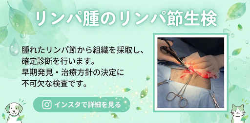
犬猫のしこりと病理検査|当院が大切にしていること
愛犬・愛猫に「しこり」や「できもの」を見つけたとき、多くの飼い主さまが一番に気になるのは「これって、がんなの?」という不安だと思います。
この記事では、当院で大切にしている「病理検査」の考え方と、より正確な診断のために行っている工夫を、できるだけ分かりやすくまとめました。
難しい専門用語はできるだけ減らしつつ、内容は省略せずにしっかりお伝えしますので、ゆっくり読んでいただけたら嬉しいです。
病理検査ってどんな検査?
- 病理検査とは、しこりや臓器から採取した細胞や組織を顕微鏡で観察し、「どんな細胞でできた病変なのか」「良性か悪性(がん)か」「治療や予後に関わる特徴はあるか」を調べる検査です。
- 細胞診(さいぼうしん)
細い針でしこりをチクッと刺して、中の細胞を少しだけ採取し、スライドガラスに塗って顕微鏡で見る方法です。
・体への負担が少なく、外来で短時間に行えることが多い
・「ざっくりとしたタイプ」を知るのが得意で、リンパ腫や肥満細胞腫などの診断に役立ちます。 - 組織検査(生検・せいけん)
しこりの一部または全部を小さく切り取って、薄く切った標本を詳しく調べる方法です。
・全体の構造や周りとの境目、細胞の並び方など、より多くの情報が得られます。
・局所麻酔や全身麻酔、手術が必要になることもありますが、治療方針を決めるうえでとても大切な検査です。 - 病理検査で分かること
・良性か悪性かの判断
・腫瘍の種類(リンパ腫・肥満細胞腫・腺がん・肉腫 など)
・増殖の勢い(悪性度)や、今後の再発・転移リスクの見通し
・場合によっては、その後に使えるお薬の種類や追加検査の必要性 など
検体の「準備」が大切な理由
- どんなに腕の良い病理医でも、送られてきたサンプルが不十分だと、診断がむずかしくなってしまいます。
- たとえば、しこりのごく一部だけに針が入って、たまたま腫瘍細胞があまり含まれていない部分しか採れていなかったり、細胞がつぶれて形が分からない状態になってしまったりすると、「はっきりとした診断名」がつけられないことがあります。
- また、固定の仕方や標本の作り方によって、細胞が本来と少し違う姿に見えてしまうこともあります。そのため、「どう採って、どう送るか」も検査の一部だと考えています。
- 当院では、病理の先生に少しでも情報を多くお届けできるよう、検体の準備と情報の添付をとても大切にしています。
当院で行っている3つの工夫
- ① しこりの「肉眼写真」を一緒に送る
手術中や診察時に、しこりの写真を撮影し、病理検査の依頼書と一緒にお送りします。
・身体のどの場所にあるのか
・表面の色や質感、周りの組織とのつながり方
などが分かることで、病理医は「顕微鏡で見えている像」と「実際の見た目」を結びつけて判断しやすくなり、診断の精度が高まりやすくなります。 - ② 組織検査と同時に、細胞診の標本も送る
特にリンパ腫や肥満細胞腫などの円形細胞腫瘍では、組織検査だけよりも、事前に行った細胞診のスライド標本があるほうが、腫瘍のタイプや悪性度を判定しやすいとされています。
そのため当院では、
・手術でとった組織の標本
・外来で採取した細胞診のスライド(必要に応じて未固定のものも含む)
をセットで病理医に送り、両方の情報を合わせて判断してもらうようにしています。 - ③ 将来の追加検査に備えた標本を用意する
腫瘍の種類によっては、後から特殊染色や免疫染色という追加検査を行うことで、より正確な診断やサブタイプ分けができる場合があります。
そこで、あらかじめ追加検査に使いやすいよう、アルコールで固定していない塗抹標本などを別に用意し、必要なときにスムーズに追加検査を依頼できるようにしています。
それでも診断がはっきりしないことがある理由
- 病理検査は「必ず一回で何でも分かる魔法の検査」ではありません。
- 採取した場所にたまたま腫瘍細胞が少なかったり、壊死や強い炎症が重なっていて細胞の形が分かりにくかったりする場合、病理医が慎重に判断しても「断定はむずかしい」「〜が疑われる」といった表現になることがあります。
- とてもまれな腫瘍だったり、腫瘍と炎症性の病変が入り混じっていたりすると、「追加の特殊染色」や「再度の生検」が必要になることもあります。
- そのようなとき当院では、
・再度のサンプル採取(必要に応じて場所を変える)
・エコーやCTなどの画像検査
・他施設や専門病理医へのセカンドオピニオン
などを組み合わせて、なるべく診断の方向性をはっきりさせていくようにしています。 - 「結果がグレーで不安…」というときこそ、次の一歩を一緒に考えることが大切だと考えています。
最近のトピックス・アップデート
- 世界的にも、腫瘍の病理検査を依頼する際には「臨床情報と写真をしっかり添えること」が推奨されており、当院のように肉眼写真や詳しい経過を一緒に送るスタイルが増えています。
- 細胞診に関しても、
・薄く広く塗る
・複数の採取方法を組み合わせる
など、標本の質を高めるためのガイドラインが整ってきており、より精度の高い診断を目指す流れが進んでいます。 - さらに近年は、病理標本をデジタル化してコンピュータで解析したり、AI(人工知能)が細胞の形を自動で読み取る研究も進んでいます。今後、診断のばらつきが少なくなったり、悪性度の予測がより客観的にできるようになることが期待されています。
飼い主さまに知っておいてほしいこと
しこりや腫瘍が見つかったとき、「しばらく様子を見てから…」と思われる方も多いのですが、病理検査は「早めに行うこと」がとても大切です。小さいうちのほうが採取しやすく、検体の質も保ちやすいため、診断がつきやすくなります。
また、検査の際には、ワンちゃん・ネコちゃんの性格や体調によって、鎮静や全身麻酔が必要になることがあります。これは、痛みやストレスをできるだけ減らし、安全に、そしてきちんとしたサンプルを採るためのもので、「無理に押さえつけて採るよりも、結果的に体に優しい選択」になることも少なくありません。
「麻酔がこわい」「本当に検査は必要?」「もしがんだったらどうしよう」――そんなお気持ちは、どれも自然なものです。不安や疑問があれば、どんな小さなことでも構いませんので、診察のときに遠慮なくお話しください。
まとめ
- 病理検査は、しこりや腫瘍の「正体」や「今後の見通し」を知るための、とても大切な検査です。
- 検体の準備や情報の共有の仕方によって、診断の精度が変わることがあります。当院では、写真や細胞診標本の同送、追加検査に備えた標本の準備など、できるだけ多くの情報を病理医に届ける工夫をしています。
When you find a lump or bump on your dog or cat, the first thing that often comes to mind is, “Could this be cancer?” It’s completely natural to feel worried.
In this article, we would like to explain how we think about pathology examinations at our clinic, and the small but important steps we take to make the diagnosis as accurate as possible.
We have tried to avoid overly technical terms while still keeping all the important content, so please feel free to read through at your own pace.
What is a pathology examination?
- A pathology examination means taking cells or tissue from a lump or organ, looking at them under a microscope, and checking
“What kind of cells are forming this lesion?”, “Is it benign or malignant (cancer)?”, and “Are there features that affect treatment or prognosis?”. - Cytology (fine needle aspiration)
A thin needle is gently inserted into the lump, a small amount of cells is collected, and the material is spread on a glass slide to be examined under a microscope.
・This procedure is minimally invasive and can often be done quickly during a regular visit.
・It is especially useful for getting an overall idea of the “type” of lesion, and it helps diagnose diseases such as lymphoma and mast cell tumor. - Histopathology (biopsy)
A small piece or all of the lump is surgically removed and processed into very thin sections for detailed examination.
・It provides more information about the overall structure, the border with surrounding tissues, and the arrangement of the cells.
・Local or general anesthesia and surgery may be required, but it is a very important test for deciding on the most appropriate treatment plan. - What pathology can tell us
・Whether a lesion is benign or malignant
・The type of tumor (lymphoma, mast cell tumor, adenocarcinoma, sarcoma, etc.)
・How aggressive the tumor is (its malignancy grade) and the risk of recurrence or metastasis in the future
・In some cases, what kinds of medications might be useful and whether additional tests will be helpful
Why is “sample preparation” so important?
- Even the most skilled pathologist will have difficulty making a clear diagnosis if the sample that arrives at the laboratory is not adequate.
- For example, if the needle happens to collect tissue from an area where there are very few tumor cells, or if the cells are crushed and their shape can no longer be recognized, it may not be possible to give a firm diagnosis.
- Also, depending on how the tissue is fixed and how the slide is prepared, the cells may look slightly different from how they looked in the body. Because of this, we think of “how we collect and how we send the sample” as an essential part of the test itself.
- At our clinic, we put a lot of effort into how we prepare samples and how we provide information, so that the pathologist can receive as much useful detail as possible.
Three things we do to improve diagnostic accuracy
- 1) Sending “gross appearance” photos together with the sample
We take photos of the lump during surgery or examination and send them along with the pathology request form.
・Which part of the body the lesion is in
・The color and texture of the surface, and how it is connected to the surrounding tissue
When the pathologist can see these points, it becomes easier to link the microscopic image to the real-life appearance of the lesion, which often leads to more accurate diagnosis. - 2) Sending cytology slides together with the biopsy sample
Especially for round cell tumors such as lymphoma and mast cell tumor, having cytology slides in addition to a biopsy sample makes it easier to determine the tumor type and malignancy grade.
Therefore, at our clinic we usually send:
・The histopathology sample obtained during surgery
・Cytology slides collected beforehand in the consultation room (including unfixed smears if needed)
as a set to the pathologist, so that both sources of information can be reviewed together. - 3) Preparing extra slides in case additional tests are needed later
Depending on the disease, additional tests such as special stains or immunohistochemistry can help refine the diagnosis or classify the tumor more precisely.
To make this possible, we prepare extra smears (for example, air-dried slides that are not fixed in alcohol) in advance so that additional tests can be requested smoothly whenever they become necessary.
Why results are sometimes still “uncertain”
- Pathology is a powerful tool, but it is not a magical examination that always reveals everything in a single try.
- If the collected area happens to contain few tumor cells, or if the tissue is largely necrotic or severely inflamed, the cell shapes can be very hard to interpret. Even with careful review, the pathologist may have to say “a definite diagnosis is difficult” or “findings are suggestive of…”.
- When the tumor type is extremely rare, or when tumor and inflammatory tissue are mixed together, repeat biopsy or additional special stains may be required.
- In such situations, we may combine several approaches, such as:
・Sampling again from a different area of the lesion
・Performing imaging studies like ultrasound or CT
・Seeking a second opinion from another laboratory or specialist pathologist
to clarify the diagnosis as much as possible. - When the report feels “gray” and uncertain, that is exactly the time when we believe it is most important to think about the next step together with you.
Recent trends and updates in pathology
- Around the world, it is now strongly recommended to attach as much clinical information and photographs as possible when requesting tumor pathology, so the style of sending photos and detailed histories – like we do at our clinic – is becoming more common.
- For cytology, there are now more detailed guidelines on how to improve slide quality, such as:
・Spreading the material thinly and evenly
・Using several collection techniques for one lesion
These efforts aim to obtain clearer slides and more reliable diagnoses. - In addition, research is progressing on digital pathology, where slides are scanned and analyzed by computer, and on using AI (artificial intelligence) to automatically read cell shapes. In the future, this may help reduce differences between pathologists and allow more objective prediction of tumor behavior.
What we would like owners to know
When a lump or tumor is discovered, it is very common to feel like “Maybe I’ll just watch it for a while first…”. However, doing a pathology examination sooner rather than later is extremely important. When the lesion is still small, it is easier to sample and easier to maintain good sample quality, which means the chances of a clear diagnosis are higher.
Depending on your pet’s personality and health condition, we may recommend sedation or general anesthesia for sampling. This is not to “do something scary”, but rather to minimize pain and stress and to collect the sample safely and accurately. In many cases, this is actually a kinder choice than trying to restrain a frightened animal and take a sample while it is fully awake.
It is completely natural to feel anxious about things like “I’m worried about anesthesia”, “Do we really need this test?”, or “What if it is cancer?”. If you have any concerns or questions – no matter how small they may seem – please feel free to talk to us during your visit.
In summary
- Pathology examinations are essential to understanding the true nature and future outlook of lumps and tumors.
- The way we prepare samples and share information can have a major impact on diagnostic accuracy. At our clinic, we try to support the pathologist by sending photographs, cytology slides and extra smears for possible additional tests, so that they can see as complete a picture as possible.
当你在爱犬或爱猫身上摸到一个小包块时,很多人的第一个念头都是:“这会不会是肿瘤?”有这样的担心是非常正常的。
在这篇文章里,我们想介绍本院对“病理检查”的看法,以及为了获得尽可能准确的结果,我们在日常诊疗中做的一些小小努力。
我们尽量减少晦涩的专业术语,但不会删减重要内容,希望您可以轻松地了解病理检查在治疗中的意义。
什么是病理检查?
- 病理检查是指从包块或器官中取出细胞或组织,在显微镜下观察,判断:
“这个病变是由什么样的细胞组成?”、“是良性还是恶性(肿瘤)?”、“有没有会影响治疗方式和预后的信息?”等。 - 细胞学检查(细针穿刺)
用一根很细的针轻轻刺入包块,吸取少量细胞,涂在玻片上,在显微镜下观察。
・对身体的负担较小,很多时候可以在门诊很快完成。
・比较擅长了解“病变的大致类型”,对淋巴瘤、肥大细胞瘤等疾病的诊断很有帮助。 - 组织病理检查(活检)
把包块的一部分或全部切除,制成很薄的切片,在显微镜下做更详细的观察。
・可以看到整体结构、与周围组织的边界、细胞的排列方式等更多信息。
・有时需要局麻或全麻以及手术,但在制定治疗方案时是一项非常重要的检查。 - 病理检查能告诉我们什么
・病变是良性还是恶性
・肿瘤的种类(如淋巴瘤、肥大细胞瘤、腺癌、肉瘤等)
・肿瘤的恶性程度、将来复发或转移的风险
・在某些情况下,还能提示可以使用的药物类型以及是否需要追加检查
为什么“样本准备”这么重要?
- 即使是经验非常丰富的病理医生,如果送去的样本质量不好,也很难做出明确的诊断。
- 比如,针刚好扎在肿瘤细胞比较少的地方,或者细胞在制片过程中被压碎、形态模糊不清,即使认真查看,也很难得到清晰的结论。
- 另外,根据固定方式和制片方法的不同,细胞在玻片上的样子有时会与在体内时略有差别。因此,我们认为,“如何取样、如何寄送”本身就是病理检查的一部分。
- 在本院,我们非常重视样本的处理和随附信息的填写,希望尽可能把有用的信息完整地传达给病理医生。
为了提高诊断准确度,我们做的三件事
- ① 与标本一起提供“肉眼所见”的照片
在手术或门诊检查时,我们会拍摄包块的照片,并与病理申请单一同寄出。
・病变位于身体的哪个部位
・表面的颜色、质地,与周围组织的连接方式
这些信息能够帮助病理医生把显微镜下看到的图像与实际样子联系起来,从而更容易做出准确判断。 - ② 组织标本和细胞学玻片同时送检
对于淋巴瘤、肥大细胞瘤等圆形细胞肿瘤,仅有组织切片时有时难以判断具体类型和恶性程度,而如果同时有细胞学玻片,会大大提高诊断的把握度。
因此,我们通常会把:
・手术中取到的组织标本
・门诊中事先做好的细胞学玻片(必要时包括未固定的风干涂片)
成套寄给病理医生,让他们将两方面的信息综合起来判断。 - ③ 预留可用于追加检查的标本
对于某些肿瘤,追加特殊染色或免疫组化染色可以帮助进一步明确诊断或进行更细致的分型。
为了在需要时能够顺利进行这些检查,我们会事先准备一些额外的涂片,比如未用酒精固定的风干涂片等,以便在日后根据需要追加检测。
即使这样,有时结果仍然“不够明确”的原因
- 病理检查非常重要,但它并不是“做一次就什么都能知道的魔法检查”。
- 如果取样部位肿瘤细胞很少,或者组织大部分已经坏死、伴有剧烈炎症,细胞形态就会变得很难辨认。即使病理医生非常谨慎,有时也只能写出“难以做出确定诊断”或“提示为……可能性较大”等描述。
- 当肿瘤类型十分罕见,或者肿瘤组织与炎症组织混在一起时,可能需要进行再次活检或追加特殊染色等检查。
- 在这种情况下,我们通常会综合采用多种方式,例如:
・从不同部位重新取样
・进行超声、CT 等影像学检查
・向其他实验室或专科病理医生寻求第二意见
尽量把诊断方向弄得更清楚。 - 当报告看起来有些“灰色地带”时,恰恰是需要与您一起讨论下一步方案的关键时刻。
病理领域的最新动向与进展
- 目前,在全球范围内,给肿瘤送检时尽可能详细地提供临床信息和照片已被广泛推荐。像我们这样提供肉眼照片、病史和检查结果的做法,在兽医界也越来越普遍。
- 关于细胞学检查,现在也有了更具体的操作指南,例如:
・尽量把涂片铺得均匀而薄
・对同一个病变采用多种取样方法
这些做法都是为了获得质量更好的玻片,从而提高诊断的可靠性。 - 此外,数字病理和人工智能也在不断发展:把玻片扫描成数字图像,通过计算机分析,甚至利用 AI 自动识别细胞形态。未来,有望减少不同病理医生之间的差异,让对肿瘤行为的预测更加客观。
希望家长了解的几点
发现包块或肿瘤时,很多人会想:“先观察一段时间再说吧……”。但事实上,越早进行病理检查越重要。在病变还比较小的时候,更容易取到合适的样本,样本质量也更容易保证,因此更有机会得到清晰的诊断结果。
根据动物的性格和身体状况,有时我们会建议在镇静或全麻下取样。这并不是为了“做更可怕的事情”,而是为了减少疼痛和压力,让取样过程更安全、更准确。很多时候,这比在动物清醒、紧张反抗时强行取样,对身体更加温和。
“我担心麻醉”、“真的有必要做这个检查吗?”、“如果真的是肿瘤怎么办?”——有这些不安都是很自然的。如果您心里有任何疑问或顾虑,无论看起来多么细小,都欢迎在就诊时随时与我们交流。
小结
- 病理检查是了解包块或肿瘤“真实身份”和“未来走向”的关键步骤。
- 样本的准备方式以及信息的传达方式会直接影响诊断结果的准确性。本院通过提供照片、细胞学玻片以及可用于追加检查的预留标本,尽量为病理医生呈现一个更完整的病例全貌。